Definition
Uterine fibroids represent the most frequent benign lesion of the uterus and develop predominantly during reproductive age. Also known as uterine leiomyomas or myomas, they are not generally associated with an increased risk of malignant uterine tumor, and only exceptionally are they linked to neoplastic disease.
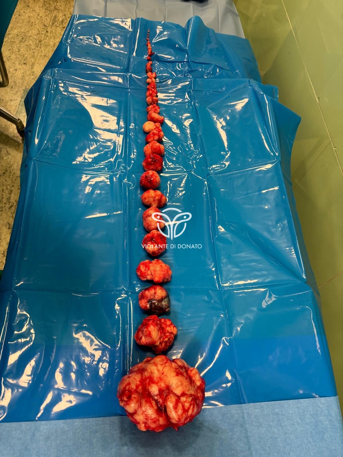
Uterine fibroid size can vary considerably: from microscopic nodules invisible to the naked eye to large masses capable of distorting the uterus and significantly increasing its volume. They may be present as single formations or as multiple lesions. In the most advanced cases, the presence of numerous fibroids may cause uterine enlargement reaching high abdominal levels.
A significant proportion of women develop uterine fibroids during their lifetime. However, many patients remain asymptomatic and the diagnosis is frequently incidental during a gynecologic examination or routine pelvic ultrasound.
Symptoms and anatomical classification
Many women with uterine fibroids do not present obvious symptoms. When symptoms are present, their intensity depends mainly on the location, number, and size of the fibroids.
In symptomatic patients, the most frequent signs include:
- Heavy menstrual bleeding
- Menstruation lasting more than one week
- Pelvic pressure or pain
- Frequent urination or difficulty in fully emptying the bladder
- Constipation
- Lower back pain or pain radiating to the lower limbs
- More rarely, acute pelvic pain associated with fibroid degeneration or torsion
Anatomical classification
From an anatomical standpoint, uterine fibroids are classified according to their site of development:
- Intramural fibroids — develop within the thickness of the uterine muscle wall (myometrium); these are the most frequent
- Submucosal fibroids — protrude into the uterine cavity; these are most frequently associated with abnormal bleeding and reproductive problems
- Subserosal fibroids — grow toward the outside of the uterus, in the direction of the abdominal cavity; they generally cause fewer hemorrhagic symptoms
FIGO has established a numbering system from 0 to 8 to define the exact location of the myoma within the uterus. This distinction is the "gold standard" for the surgeon in choosing the operative instrumentation (e.g. hysteroscopic resectoscope vs laparoscopic or robotic approach).

FIGO subclassification system for uterine leiomyomas (0–8). Red: submucosal (SM) · Green: intramural/subserosal · Yellow: hybrids
| Grade | Category | Description |
|---|---|---|
| Submucosal Myomas (SM) — Grade 0–2 | ||
| 0 | Submucosal | Completely intracavitary — pedunculated |
| 1 | Submucosal | <50% intramural (predominantly intracavitary) |
| 2 | Submucosal | ≥50% intramural |
| Other Myomas (O) — Grade 3–8 | ||
| 3 | Intramural | Contacts the endometrium; entirely intramural (100%) |
| 4 | Intramural | Purely intramural, without contact with endometrium or serosa |
| 5 | Subserosal | Subserosal ≥50% intramural |
| 6 | Subserosal | Subserosal <50% intramural |
| 7 | Subserosal | Subserosal pedunculated (completely extracavitary) |
| 8 | Other locations | Other locations: cervical, parasitic, broad ligament |
| Hybrid Myomas — e.g., 2-5 | ||
| 2-5 | Hybrid | Two numbers separated by hyphen: the first indicates the relationship with the mucosa, the second with the serosa. E.g., 2-5: ≥50% intramural on the endometrial side and ≥50% intramural on the serosal side. |
Clinical implications for the choice of approach
- Grades 0–2 (submucosal): hysteroscopic approach with resectoscope; grade 0 simpler, grade 2 requires greater experience
- Grades 3–4 (intramural): laparoscopic, mini-laparotomic, or robotic myomectomy; grade 3 may require endometrial incision
- Grades 5–7 (subserosal): laparoscopy, mini-laparotomy, or robotic; grade 7 pedunculated technically more favorable
- Grade 8: approach to be evaluated case by case
- Hybrids: dual approach (hysteroscopy + laparoscopy) often required in the same operative session
FIGO = Fédération Internationale de Gynécologie et d'Obstétrique. Int J Gynaecol Obstet. 2011;113(1):3-13.
Causes
The exact causes of uterine fibroids are not fully understood. However, several biological factors appear to contribute to the development of these lesions.
- Genetic alterations. Many fibroids show genetic alterations compared to normal smooth muscle cells of the myometrium.
- Hormones. Estrogens and progesterone promote fibroid growth. Fibroid cells express a greater number of receptors for these hormones than normal myometrial cells. After menopause, the reduction in hormonal production is associated with a decrease in fibroid size.
- Growth factors. Some molecules such as insulin-like growth factor may contribute to fibroid development and proliferation.
- Extracellular matrix (ECM). In fibroids this matrix is particularly abundant, contributing to the fibrous consistency of the lesions.
Fibroids are believed to derive from clonal proliferation of a single myometrial stem cell. Growth behavior may vary: some lesions grow slowly, others remain stable, and still others shrink spontaneously. During pregnancy some fibroids temporarily increase in size, but tend to decrease after delivery.
Risk factors
- Race. Prevalence is higher in women of African descent, who tend to develop fibroids at an earlier age, with larger size and more pronounced symptoms.
- Family history. The presence of fibroids in first-degree relatives — mother or sisters — increases the risk of developing the condition.
- Other factors. Early menarche, obesity, low vitamin D levels, a diet rich in red meat and low in fruits, vegetables, and dairy products, as well as alcohol consumption, have been associated with an increased likelihood of developing uterine fibroids.
Complications
Although uterine fibroids are generally benign, they may cause clinically relevant symptoms. The most frequent complication is iron deficiency anemia due to heavy and prolonged menstrual bleeding, which may manifest with fatigue, weakness, and reduced functional capacity. Only rarely is the blood loss sufficient to require transfusion.
Malignant transformation of a benign fibroid into leiomyosarcoma is extremely rare. Studies report a prevalence of occult leiomyosarcoma in women operated for fibroids ranging from approximately 1 in 352 to 1 in 8,300 cases. Leiomyosarcomas generally represent distinct tumors that do not derive directly from fibroid degeneration.
Pregnancy and fibroids
In most cases, uterine fibroids do not prevent conception. However, certain locations, particularly submucosal fibroids, may interfere with embryonic implantation and increase the risk of infertility or miscarriage.
During pregnancy, the presence of fibroids may be associated with an increased risk of certain obstetric complications, including placental abruption, fetal growth restriction, and preterm birth. Assessment of these risks requires individualized specialist evaluation.
Initial diagnostic algorithm — Suspected uterine fibroids
(heavy bleeding, compression symptoms, pelvic pain, reproductive problems)
Therapeutic algorithm — Symptomatic fibroids
Adapted from: Uterine Fibroids. N Engl J Med. 2024;391(18):1721-1733.
Treatment
Treatment of uterine fibroids must be personalized and depends on symptom severity, lesion size and location, patient age, and pregnancy desire.
Clinical observation
In cases where fibroids are small, asymptomatic, or do not cause clinically relevant consequences, periodic monitoring with clinical and ultrasound follow-up may be indicated. This approach is frequently adopted in patients approaching menopause.
Pharmacologic treatment
Some hormonal therapies may contribute to menstrual cycle regulation and bleeding reduction. GnRH agonists cause temporary hormonal suppression and may reduce fibroid size, generally as preoperative treatment. Oral GnRH antagonists may significantly reduce bleeding. Hormonal contraceptives and levonorgestrel intrauterine devices act primarily on symptom control.
Laparoscopic, mini-laparotomic, and hysteroscopic myomectomy
Myomectomy consists of selective surgical removal of fibroids with uterine preservation. It can be performed via laparoscopic, mini-laparotomic, or hysteroscopic approach depending on the location and size of the lesions. This procedure is indicated in women who wish to preserve fertility or retain the uterus. Laparoscopic and mini-laparotomic myomectomy are associated with reduced blood loss, shorter hospital stay, and faster recovery compared to traditional laparotomic surgery.
Uterine artery embolization
Uterine artery embolization (UAE) is a non-surgical procedure performed in interventional radiology that reduces blood supply to fibroids, leading to volume reduction. It requires multidisciplinary evaluation with the gynecologist and interventional radiologist.
Hysterectomy
In the most complex cases or when conservative therapies are ineffective, removal of the uterus may be indicated. This is a definitive treatment that eliminates the risk of recurrence, but it is not appropriate for women who wish to have a future pregnancy.
Prevention
Current scientific knowledge does not allow identification of definitive preventive strategies for uterine fibroids. However, certain lifestyle habits may contribute to risk reduction: maintaining adequate body weight and a diet rich in fruits and vegetables appear to have a possible protective effect. Some studies suggest that the use of hormonal contraceptives may be associated with a lower incidence of uterine fibroids.
- Persistent pelvic pain
- Very heavy, prolonged, or painful menstruation
- Bleeding between cycles
- Difficulty emptying the bladder
- Anemia symptoms (fatigue, paleness, weakness)
- Significant vaginal bleeding or sudden-onset acute pelvic pain — urgent evaluation
Frequently asked questions
Fibroid, myoma, and leiomyoma indicate the same benign formation of the uterus originating from the smooth muscle cells of the myometrium. The three terms are interchangeable in clinical practice. The term fibromatous uterus is used when multiple fibroids are present in the uterus simultaneously.
Ref.: [7, 16, 20]
Malignant transformation of a benign fibroid into leiomyosarcoma is extremely rare. Studies report that the prevalence of occult leiomyosarcoma in women operated for suspicious fibroids ranges from approximately 1 in 352 to 1 in 8,300 cases. Leiomyosarcomas generally represent distinct tumors that do not derive directly from the degeneration of benign fibroids.
Ref.: [4, 5, 6]
Surgery is indicated when:
- Symptoms (heavy bleeding, pelvic pain, compression symptoms) significantly compromise quality of life
- Anemia from chronic bleeding develops
- Fibroids cause infertility or obstetric complications
- The patient desires a definitive solution
Monitoring is appropriate when:
- Fibroids are asymptomatic
- The patient does not want intervention
- The patient is in perimenopause (fibroids tend to regress after menopause)
Ref.: [7, 14, 16, 18]
The impact of fibroids on fertility depends primarily on their location. Submucosal fibroids are most frequently associated with infertility and miscarriage, as they may interfere with embryonic implantation and alter endometrial vascularization. Intramural fibroids may reduce fertility in some patients, while subserosal fibroids generally have little impact on reproductive capacity.
During pregnancy they may increase the risk of obstetric complications such as preterm birth, placental abruption, or fetal growth restriction.
Ref.: [8, 9, 10, 11]
Laparoscopic and mini-laparotomic myomectomy are associated with reduced intraoperative blood loss, shorter hospital stay, faster functional recovery, and less postoperative pain. Numerous studies and meta-analyses also demonstrate a reduction in complications compared to traditional laparotomic surgery.
Mini-laparotomy (4–7 cm incision) represents a valid alternative to laparoscopy, particularly for large fibroids or in cases of complex uterine anatomy, with comparable clinical outcomes and a more accessible learning curve.
However, situations remain in which traditional laparotomy may be preferable, for example in the presence of very voluminous fibroids, unfavorable location, or severe uterine anatomical distortion. The choice of approach must be personalized by the surgeon based on individual characteristics.
Ref.: [1, 12, 13, 18]
Some medications may temporarily reduce fibroid volume and control symptoms. GnRH agonists may cause uterine volume reduction of up to 30–60% but are generally used for limited periods (3–6 months) as preoperative treatment, due to side effects from hypoestrogenism.
Oral GnRH antagonists (relugolix, elagolix) may significantly reduce bleeding with a more manageable side-effect profile. Hormonal contraceptives or levonorgestrel intrauterine devices act primarily on symptom control without reducing fibroid size.
Ref.: [7, 14, 15, 16]
After myomectomy, the recurrence risk is not negligible. Studies indicate fibroid recurrence in 25% of cases within approximately 40 months and in 23–30% of cases within 5–7 years. The risk is greater in younger patients, in the presence of multiple fibroids, and in cases where the primary fibroid has not been completely removed.
For this reason, planning the timing of surgery — in relation to the patient's age and reproductive desire — is a central element of specialist consultation.
Ref.: [7, 17]
Hysterectomy represents the definitive treatment because it completely eliminates the uterus and therefore the risk of fibroid recurrence. However, it is not the only therapeutic option. In many patients, conservative strategies such as myomectomy, ablative techniques, and other procedures may be considered, depending on clinical characteristics and reproductive desires.
The decision must be shared between the patient and the surgeon, taking into account symptoms, age, fertility desire, and the anatomical characteristics of the fibroids.
Ref.: [7, 14, 18]
With menopause, there is a reduction in estrogen and progesterone production, hormones that stimulate fibroid growth. As a result, many fibroids tend to shrink spontaneously and symptoms — particularly heavy menstrual bleeding — resolve with cessation of menstruation.
If a fibroid continues to grow after menopause, urgent specialist evaluation is indicated, as this behavior is atypical and must rule out malignant pathology.
Ref.: [7, 16, 19]
Specialist Consultation Request
For evaluation of uterine fibroids, to discuss the indication for laparoscopic, mini-laparotomic, or hysteroscopic myomectomy, or for a surgical second opinion, you may request a consultation with Prof. Di Donato.
Book a consultation →